Digital Topography
Digital Topography
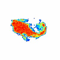
Topography is the high spatial resolution study of an individual reflection. It reveals the mosaic structure of the crystal. Normally the images are captured with high resolution nuclear emulsion plates. This process is time consuming and complicated. It can take a long time to acquire complete data for a single reflection.
We have replaced the film with a CCD. We use direct conversion of the X-rays at the surface of the CCD to collect the data. The CCD does not have the same high resolution of the nuclear emulsion plates but it is much faster and easier to use with almost comparable resolution.
Initial Results
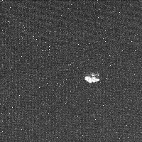
First topograph recorded at NSLS Feburary 2002.
Room Temperature Results
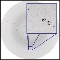
MarCCD coarse image with area of topography collection exploded.
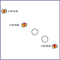
Summation of topographs for each reflection. Reflections are shown approximately 2x normal size.
Cryo Results
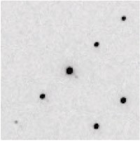
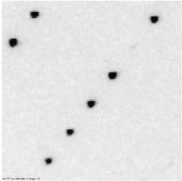
Two regions of a cryogenically frozen crystal of lysozyme where digital topographs were taken of size reflections in each region.
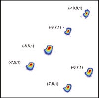
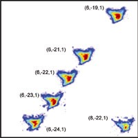
Summation of topographs for each reflection. Reflections are shown approximately 2x normal size.
Fine-Phi Crystal Studies
Fine-Phi Crystal Studies
Earth-Grown Insulin
Earth-Grown Insulin
Shown are two different reflections that were sampled every 0.001 degrees for a total of one degree. Below is the corresponding coarse oscillation image with the predicted reflection locations overlaid with red circles. Reflections overlaid in yellow circles failed to pass a series of filters designed to find usable reflection profiles that were accurately recorded. Reflections with black and green crosshairs are further analyzed below.
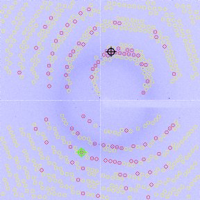
Coarse (1.0°) Oscillation Image From an Earth-grown Insulin Crystal
Topography/Reflection Profile/3D Reflection Animations
For each reflection, the following comparisons are made (From left to right): (1) coarse image (1.0°), (2) topographs and corresponding reflection profile from the super fine phi (0.001°) slicing technique, and (3) 3-D reconstruction from the super fine phi images (0.001°) with x and y detector pixel positions and z phi angle in degrees.
Reflection H=-20 K=1 L=10 (Black)

Coarse Image
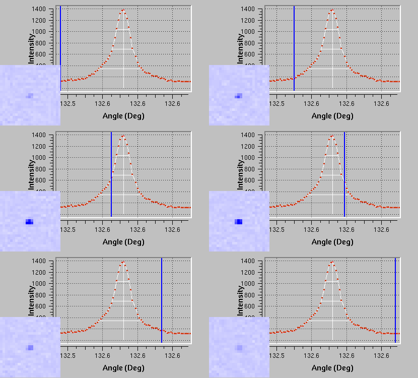
Topograph/Reflection Profile (0.001° sampling)
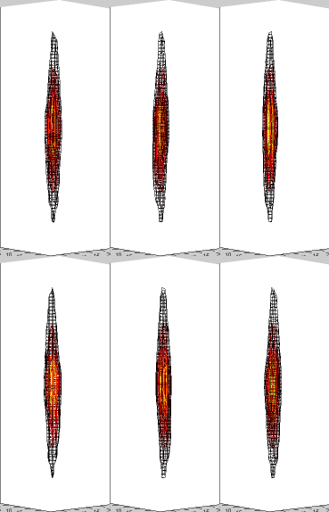
3D Reflection Rotation
(rotation about vertical phi axis)
Reflection H=16 K=7 L=-3 (Green)

Coarse Image
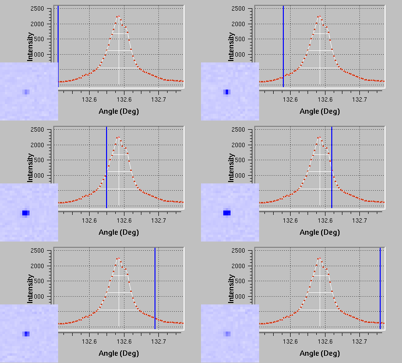
Topograph/Reflection Profile (0.001° sampling)
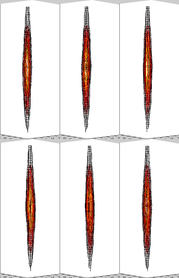
3D Reflection Rotation
(rotation about vertical phi axis)
Microgravity-Grown Insulin
Microgravity-Grown Insulin
Shown are two different reflections that were sampled every 0.001 degrees for a total of one degree. Below is the corresponding coarse oscillation image with the predicted reflection locations overlaid with red circles. Reflections overlaid in yellow circles failed to pass a series of filters designed to find usable reflection profiles that were accurately recorded. Reflections with black and green crosshairs are further analyzed below.
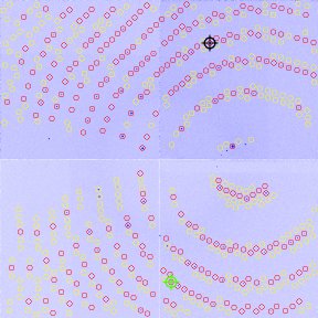
Coarse (1.0°) Oscillation Image From a Microgravity-grown Insulin Crystal
Topography/Reflection Profile/3D Reflection Animations
For each reflection, the following comparisons are made (From left to right): (1) coarse image (1.0°), (2) topographs and corresponding reflection profile from the super fine phi (0.001°) slicing technique, and (3) 3-D reconstruction from the super fine phi images (0.001°) with x and y detector pixel positions and z phi angle in degrees.
Reflection H=1 K=6 L=12 (Black)

Coarse Image
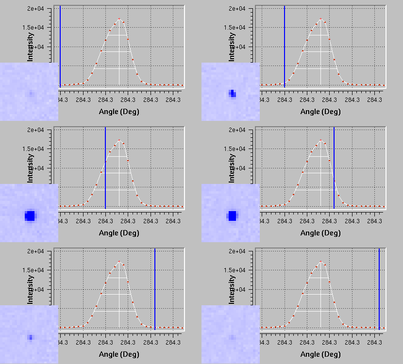
Topograph/Reflection Profile (0.001° sampling)
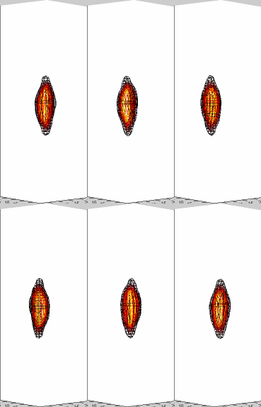
3D Reflection Rotation
(rotation about vertical phi axis)
Reflection H=-18 K=32 L=-2 (Green)

Coarse Image
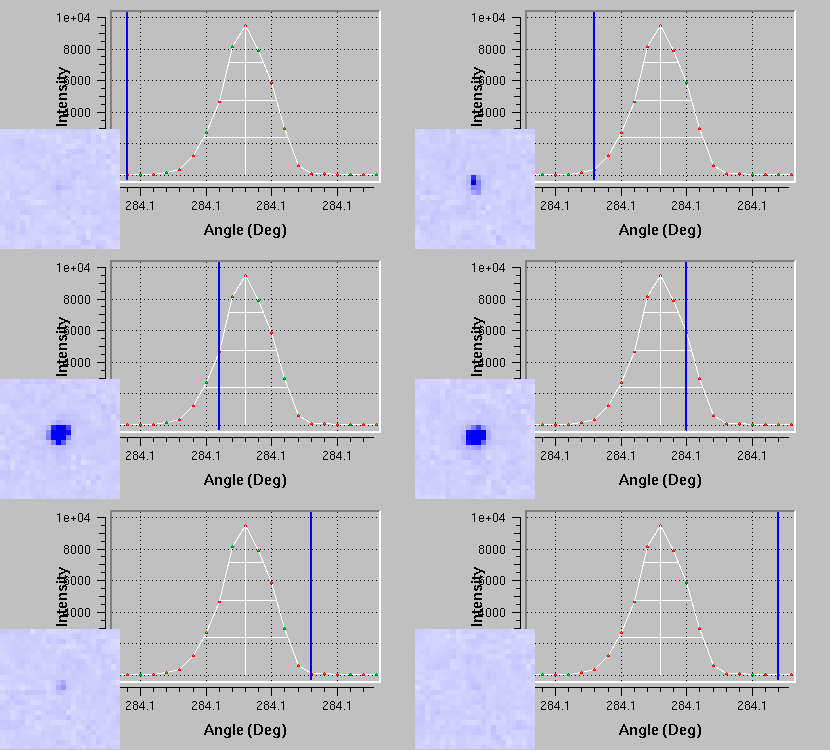
Topograph/Reflection Profile (0.001° sampling)

3D Reflection Rotation
(rotation about vertical phi axis)
Actin:Profilin
Actin:Profilin
Shown are three different reflections that were sampled every 0.01 degrees for a total of five degrees. Below is the corresponding coarse oscillation image with the predicted reflection locations overlaid with red circles. Reflections overlaid in yellow circles failed to pass a series of filters designed to find usable reflection profiles that were accurately recorded. Reflections with black and green crosshairs are further analyzed below.

Coarse (5.0°) Oscillation Image From a Microgravity-grown Insulin Crystal
Topography/Reflection Profile/3D Reflection Animations
For each reflection, the following comparisons are made (From left to right): (1) coarse image (5.0°), (2) topographs and corresponding reflection profile from the super fine phi (0.01°) slicing technique, and (3) 3-D reconstruction from the super fine phi images (0.01°) with x and y detector pixel positions and z phi angle in degrees.
Reflection H=1 K=6 L=12 (Yellow)
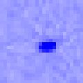
Coarse Image
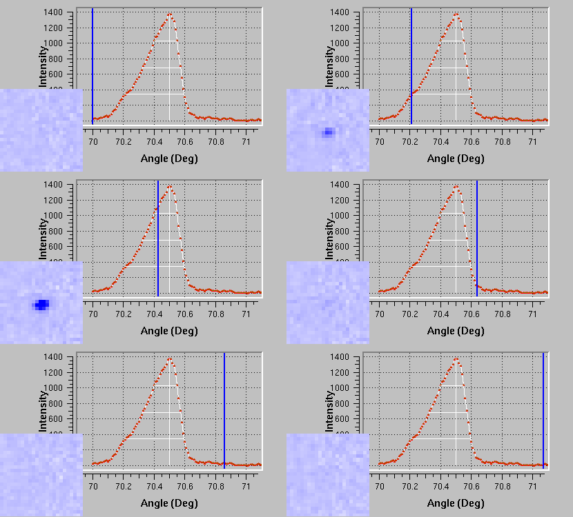
Topograph/Reflection Profile (0.01° sampling)

3D Reflection Rotation
(rotation about vertical phi axis)
Reflection H=-2 K=4 L=21 (Green)

Coarse Image
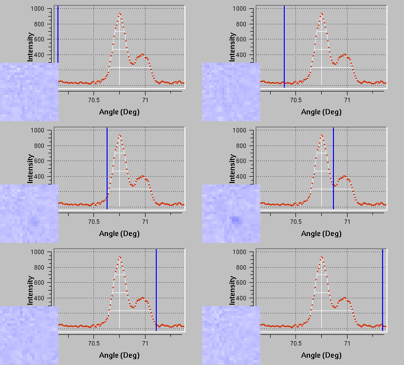
Topograph/Reflection Profile (0.01° sampling)

3D Reflection Rotation
(rotation about vertical phi axis)
Reflection H=3 K=-1 L=-38 (Black)

Coarse Image
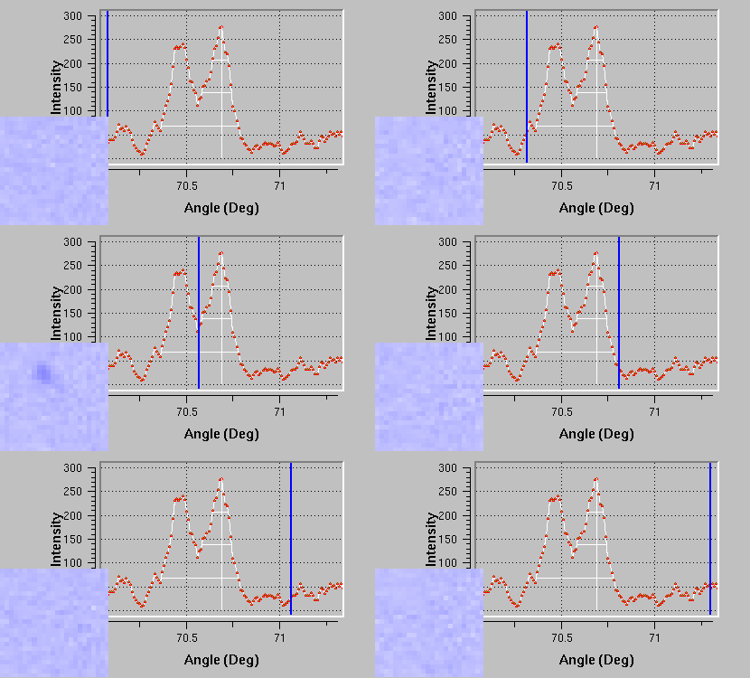
Topograph/Reflection Profile (0.01° sampling)

3D Reflection Rotation
(rotation about vertical phi axis)
